Home
/ External Nose Anatomy : Cunningham S Text Book Of Anatomy Anatomy Fig 669 Profile Yizvr Of The Bont And Carti Laginous Skeleton Of The External Nose Xasal Bone Frontal Process Of Maxilla Lateral Cartilage Fig 670 Front View : Rhinoplasty involves manipulating, shaving, adding to and/or otherwise altering the structures of the nose.
External Nose Anatomy : Cunningham S Text Book Of Anatomy Anatomy Fig 669 Profile Yizvr Of The Bont And Carti Laginous Skeleton Of The External Nose Xasal Bone Frontal Process Of Maxilla Lateral Cartilage Fig 670 Front View : Rhinoplasty involves manipulating, shaving, adding to and/or otherwise altering the structures of the nose.
External Nose Anatomy : Cunningham S Text Book Of Anatomy Anatomy Fig 669 Profile Yizvr Of The Bont And Carti Laginous Skeleton Of The External Nose Xasal Bone Frontal Process Of Maxilla Lateral Cartilage Fig 670 Front View : Rhinoplasty involves manipulating, shaving, adding to and/or otherwise altering the structures of the nose.. The lower cartilage adds support, width, and height. An area called the choana links the nasopharanx to the left and right portions of the nasal cavity, which is the space above the oral cavity and hard palate. Your nose also has four types of muscles called elevators, depressors, compressors and dilators. In the lateral view of the skeleton, the uppermost part of the nasal bone connects to the frontal bone by the nasofrontal suture. To increase the size of the nose or provide more structural support, grafts can be made from existing cartilage in the patient's nasal septum, the ear, or rib cage.
The anterior ethmoidal artery, which is also a branch of the ophthalmic artery, emerges from the base between the nasal bone and the lateral nasal cartilage and runs downward to the nasal tip. The root is continuous with the anterior surface of the head and the part between the root and the apex is called the dorsum of the nose. The septum is comprised of bone in the back and cartilage in the middle and front portions. See full list on plasticsurgerykey.com The entire nasal cavity is lined with a mucosal surface made up of epithelial cells and glands that produce mucus.
The Nose And Sinuses Teachmeanatomy from teachmeanatomy.info See full list on plasticsurgerykey.com In the lateral view of the skeleton, the uppermost part of the nasal bone connects to the frontal bone by the nasofrontal suture. Ex·ter·nal nose the visible portion of the nose that forms a prominent feature of the face; Located below the skull base and intracranial compartment, it is divided by the septum. Bone supports the upper third (bridge) of the nose. More images for external nose anatomy » A basal portion made up of the columella, nostrils, soft tissues and infra tip lobule; Figure 5 shows the basic components of the nose that are altered during rhinoplasty procedures.
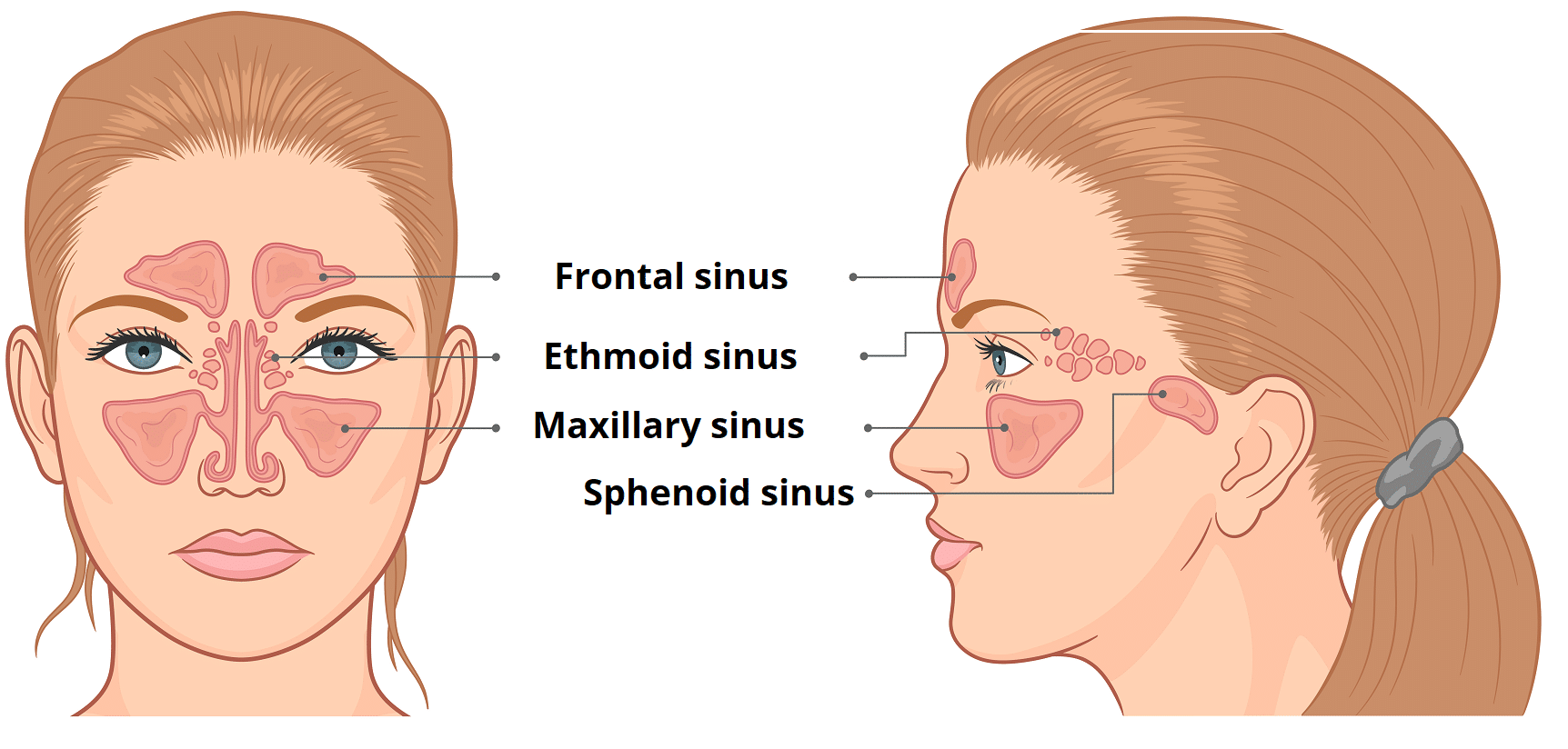
The middle turbinate is located next to the septum and extends into the central nasal cavity.
Four arteries originating either from the internal carotid or from the external carotid arteries are the main supply for the external nose (table 17.2). It is attached to the sidewall at the back, right above the inferior turbinate. Sep 29, 2019 · it extends from the vestibule of the nose to the nasopharynx, and has three divisions: It provides the support for the upper portion of the nose. And two other portions called the latter and oblique sections. Below the septal cartilage is the greater alar cartilage, which forms the lateral and medial walls of the nostrils, and below this are four lesser alar cartilages. To decrease the size of the nose in any specific area, cartilage can be strategically sutured without sacrificing the support structure. Together, the greater alar cartilage and the lesser alar cartilages give your tip and nostrils their shape. Pairs of nasal bones form the upper part and frontal processes of the maxilla, the middle part is made of pairs of upper lateral cartilages, and the lower part consists of lower lateral cartilages. At the very top of the nasal anatomy is the nasopharanx, which contains lymphoid tissue known as the adenoids. May 31, 2021 · the external nasal anatomy is quite simple. The entire nasal cavity is lined with a mucosal surface made up of epithelial cells and glands that produce mucus. The root is continuous with the anterior surface of the head and the part between the root and the apex is called the dorsum of the nose.
Nose Anatomy Stock Illustrations 5 577 Nose Anatomy Stock Illustrations Vectors Clipart Dreamstime from thumbs.dreamstime.com And two other portions called the latter and oblique sections. To decrease the size of the nose in any specific area, cartilage can be strategically sutured without sacrificing the support structure. Your nose also has four types of muscles called elevators, depressors, compressors and dilators. The bony and cartilaginous frame of the nose can be divided into three parts according to structure (fig. An area called the choana links the nasopharanx to the left and right portions of the nasal cavity, which is the space above the oral cavity and hard palate. Rhinoplasty involves manipulating, shaving, adding to and/or otherwise altering the structures of the nose. The nasal bone is also fused with the frontal bone superiorly by the frontonasal suture and with the frontal process of the maxilla by the nasomaxillary suture. The turbinates regulate nasal airflow and provide mucosa surface area.
Bone supports the upper third (bridge) of the nose.
The nasal bone is also fused with the frontal bone superiorly by the frontonasal suture and with the frontal process of the maxilla by the nasomaxillary suture. To increase the size of the nose or provide more structural support, grafts can be made from existing cartilage in the patient's nasal septum, the ear, or rib cage. It is located above and behind the middle. The bony and cartilaginous frame of the nose can be divided into three parts according to structure (fig. The portion of the nostrils that attach to the cheeks is called the alae. The inferior border joins to the upper lateral cartilage. The root is continuous with the anterior surface of the head and the part between the root and the apex is called the dorsum of the nose. Four arteries originating either from the internal carotid or from the external carotid arteries are the main supply for the external nose (table 17.2). Heading downward along the nasal ridge, the long straight part protruding anteriorly is the nasal dorsum. The septum is comprised of bone in the back and cartilage in the middle and front portions. In this sense, the nose is an air filter that purifies air before it reaches the lungs. See full list on plasticsurgerykey.com See full list on plasticsurgerykey.com
The middle turbinate is located next to the septum and extends into the central nasal cavity. The nasal dorsum is narrowest at the intercanthal line, which is the line connecting the bilateral medial canthal tendons that becomes wider as you move down the nose. The lower cartilage adds support, width, and height. A basal portion made up of the columella, nostrils, soft tissues and infra tip lobule; The upper cartilage supports the side of the nose.
Nose Aarontrinidade from static.wixstatic.com The septal cartilage, which is moveable, is surrounded by another section of cartilage called the lateral nasal cartilage. Figure 5 shows the basic components of the nose that are altered during rhinoplasty procedures. Millions of cilia continually move the mucus layer across the mucosal surface, pushing harmful particles out of the nose in the process. Rhinoplasty involves manipulating, shaving, adding to and/or otherwise altering the structures of the nose. Your nose also has four types of muscles called elevators, depressors, compressors and dilators. The bony and cartilaginous frame of the nose can be divided into three parts according to structure (fig. The nasal bone is also fused with the frontal bone superiorly by the frontonasal suture and with the frontal process of the maxilla by the nasomaxillary suture. The dorsal nasal artery emerges from the orbital cavity to the subcutaneous layer above the medial canthal tendon, which runs obliquely inferomedially and distributes in the upper dorsal part of the nose.
In the lateral view of the skeleton, the uppermost part of the nasal bone connects to the frontal bone by the nasofrontal suture.
See full list on plasticsurgerykey.com The anterior ethmoidal artery, which is also a branch of the ophthalmic artery, emerges from the base between the nasal bone and the lateral nasal cartilage and runs downward to the nasal tip. May 31, 2021 · the external nasal anatomy is quite simple. The entire nasal cavity is lined with a mucosal surface made up of epithelial cells and glands that produce mucus. The septum is comprised of bone in the back and cartilage in the middle and front portions. The internal structure of the nose, namely its cartilage and bone structure, is responsible for the physical/ visible characteristics of the nose. Heading downward along the nasal ridge, the long straight part protruding anteriorly is the nasal dorsum. The junctional point between the upper lateral cartilage and lower lateral cartilage is the supratip breakpoint, which is the inferior border of the nasal dorsum. The inferior border joins to the upper lateral cartilage. The midline point on the suture is defined as the nasion. To increase the size of the nose or provide more structural support, grafts can be made from existing cartilage in the patient's nasal septum, the ear, or rib cage. The smallest of the three turbinates is called the superior turbinate. This mucus keeps the inside of the nose moist, traps allergens and other particles and helps keep the air you breathe in humid.